GLP-1 drugs have transformed weight loss by suppressing appetite. The other half of the equation — helping the body burn more energy — has received less attention.
Focusing on metabolic studies in mice, a research team at the University of Wisconsin School of Medicine and Public Health is studying a molecular pathway that they hope can be targeted to increase energy expenditure and tip metabolic balance in a healthy direction.
By studying a specialized type of energy-burning fat, called brown adipose tissue or brown fat, scientists in a team led by Andrea Galmozzi, assistant professor of medicine, identified the main source of heme — a molecule essential for these fat cells to generate heat and maintain metabolic health. The findings were recently published in Nature Metabolism.
Researchers found that stopping heme production reduces the ability of brown adipose tissue to burn energy, causing an overaccumulation of metabolites also seen in obese and diabetic patients. Biologically simulating the metabolic disease conditions allowed scientists to discover a gene they suspect could be targeted to speed up heme production, thus speeding up energy burn.
Dylan Duerre, a member of Galmozzi’s team who recently finished his graduate training in endocrinology, diabetes and metabolism and started a research position in the private sector, said that the findings hold potential for developing new treatments for diabetes and obesity.
“It has been challenging to identify appropriate strategies to increase energy expenditure at the pharmacologic level,” Duerre said. “Our work may help identify a new way to increase energy expenditure that might be relevant for obesity.”
He noted the current limitations with weight‑loss drugs, such as GLP1s, including that obese patients tend to lose more weight on these medications than diabetic obese patients. The Galmozzi team’s research could eventually help close that gap.
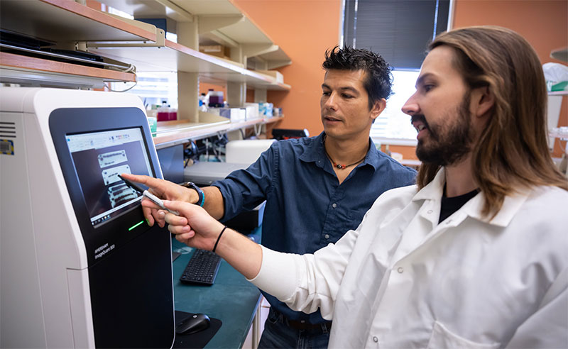
Andrea Galmozzi (left) and Dylan Duerre
“If we find a way to restore the proper expression of these enzymes, we might be able to increase the flux through this metabolic pathway, and also reduce branched-chain amino acids, and that would result in a metabolic improvement in severe disease, insulin sensitivity and glucose disorders,” Galmozzi said. “We are trying to identify some functional mechanisms that we can pharmacologically modulate to increase metabolic flux and restore metabolic illnesses.”
Diabetes and obesity are significant health issues in Wisconsin, where approximately 36% of the population has obesity and 9% has diagnosed diabetes, according to the American Diabetes Association. These diseases disproportionately affect disadvantaged populations in Wisconsin, according to the Wisconsin Department of Health Services.
Peering deep into brown fat
Brown fat gains its characteristic color by being densely packed with mitochondria, the “energy converters” of cells, which burn fuel to generate heat. Mitochondria are rich in iron-containing heme molecules, which causes the brown color that distinguishes energy-producing brown fat from energy-storing white fat.
To understand more about heme’s role in heat generation, researchers needed to know where heme was mainly being produced in brown adipose tissue, whether it was coming from inside or outside of the cells, so they tried alternatively disrupting heme production or uptake of heme.
The experiments showed that heme was primarily produced inside the brown fat cell. This allowed them to take precise genetic approaches to blocking production in brown adipose tissue.
The effect was dramatic, impacting the normal function of the tissue, which plays a specialized biological role in mammals. When internal heme production was blocked, the metabolic rate slowed, and the characteristic hue of the brown fat cells disappeared.
Some unexpected side effects
Scientists wanted to gain insight into the side effects of tampering with heme production. They set out to discover whether negative metabolic effects of blocking heme production in brown fat, which they were able to achieve in mice by deleting a gene required for heme biosynthesis, arose from the lack of heme or as a side effect of disrupting the heme production process.
To test this, scientists supplemented the fat cells of mice lacking a key heme biosynthesis gene called Alas1 with a slow-release form of heme. Supplementation restored heme levels within the cells and restored energy production, but to their surprise, thermogenesis — the ability of tissue to produce heat — was still decreased due to downregulation of Ucp1, a gene previously identified as being involved in thermoregulation.
“That discovery was probably the biggest ‘What the heck is going on?’ moment in my PhD,” Duerre said. “We had completely rescued the characteristic color of the brown adipose tissue, and they respired completely normally, but the downregulation of Ucp1 meant there had to be some really unique feature differentiating [the two pathways].”
The scientists hypothesized that an accumulation of substrates not being used up in internal heme production was causing the downregulation, rather than a lack of intracellular heme. Researchers analyzed the amount of substrates in different fat cells and found an accumulation of intermediate products, particularly those arising from branched-chain amino acids such as valine and leucine.
Think of it like a supply chain: Is the problem a lack of finished product, or a buildup of raw materials and semi-finished products from a manufacturing process gone awry?
When the heme biosynthesis process slowed down, these unused raw materials started building up, which turned Ucp1 off, reducing energy burn. Accumulation of a compound called propionyl-Coenzyme A caused a series of events that downregulated Ucp1.
A key result was that mice unable to produce heme in brown fat tissue could not tolerate cold temperatures as well as wild type mice.
“What we normally see is that on cold exposure, the BCAAs in circulation drop, and that is because they are taken up by tissues,” Galmozzi said. “In mice that lack the ability to synthesize heme in brown adipose tissue, this drop is significantly reduced — they’re not using BCAAs as they should.”
This was a major turning point for the project.
The research team concluded that heme synthesis in brown adipose tissue plays a key role in maintaining balance of branched-chain amino acid levels, linking these processes in a way that had not been identified previously.
“We’ve known that brown fat pulls a lot of branched-chain amino acids out of the bloodstream, but it wasn’t clear what it was doing with them,” Duerre said. “The big question was, where are they going? Our discovery provides, at least in part, an explanation.”
The molecular shortcut
By using BCAAs as a shortcut for heme production, brown fat conserves other nutrients, such as glucose and lipids, that can be diverted to energy production. That has the added benefit of clearing excess BCAAs from the bloodstream, which is important because accumulation of these molecules is linked to metabolic disorders.
The findings raise new questions about what happens when heme synthesis slows down or accelerates. Other researchers have previously shown that heme synthesis declines in people who are obese, but this is also true during aging, which is associated with increased risk of obesity and type 2 diabetes.
This research has been recognized for its potential impact on the field. “We found this work to be of great interest because the findings linking heme synthesis to brown adipose tissue function are unexpected yet elegantly demonstrated,” the Nature Metabolism editorial team wrote. “This study advances our understanding of adipose tissue biology and may inspire the development of new therapies targeting obesity.”
The next steps of research will focus on analyzing the effects of reduced heme production and better understanding the mechanism on a molecular level.
“We’re trying to understand whether reduction of heme production alone is sufficient to drive the metabolic defect we see in age,” Galmozzi said.
– Author Mary Bosch is a 2025–26 science communications intern at the School of Medicine and Public Health
This work was supported in part by the National Institutes of Health, including grants 1R35GM150899, P41GM108538, R35GM118110, R35GM147014, T32AG000213, TL1TR002375, UL1TR002373, and U54DK110858. Funding was also provided by the Wisconsin Partnership Program at the University of Wisconsin School of Medicine and Public Health (WPP5451), the Diabetes Research Center at Washington University (P30DK020579), the American Cancer Society (postdoctoral fellowship PF-23-1070297-01-TBE), and the American Heart Association (predoctoral fellowship 25PRE1374479).